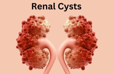


The kidneys, two bean-shaped organs nestled in the lower back, play a pivotal role in maintaining our body’s internal equilibrium. Beyond their filtration function, these remarkable organs are a fascinating testament to the complexity of human anatomy. In this article, we will delve into the anatomy of the kidney, exploring its intricate structures and functions
Basic Anatomy
Each of us possesses two kidneys, one on the left and one on the right side of the body. They are typically about the size of a human fist, though their size can vary slightly among individuals. The kidneys are positioned just below the ribcage, flanking the spine.
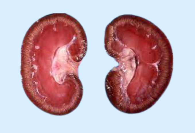
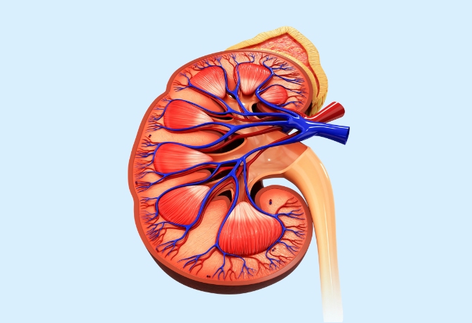
Renal Cortex and Medulla
The outermost layer of the kidney is called the renal cortex, while the inner layer is known as the renal medulla. The cortex contains millions of tiny filtering units called nephrons, which are responsible for the kidney’s primary function: filtering waste and excess substances from the blood to form urine.
Nephrons
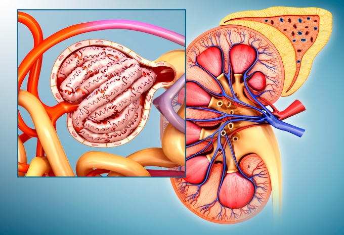
Nephrons are the kidney’s workhorses. Each kidney contains approximately one million nephrons. They consist of a renal corpuscle and a renal tubule. The renal corpuscle includes a glomerulus, a dense network of capillaries, and Bowman’s capsule, a double-walled, cup-shaped structure that encloses the glomerulus.
The renal tubule is divided into several sections, including the proximal convoluted tubule, loop of Henle, distal convoluted tubule, and collecting duct. As blood enters the glomerulus, it undergoes filtration, allowing small molecules like water, glucose, and electrolytes to pass into the renal tubule while retaining larger substances like blood cells and proteins.
Renal Arteries and Veins
Blood enters and exits the kidneys through the renal arteries and veins. The renal arteries branch off from the aorta, supplying oxygenated blood to the kidneys. After filtration, the cleansed blood returns to circulation through the renal veins, heading towards the inferior vena cava.
Ureters and Renal Pelvis
Once urine is formed within the nephrons, it flows through a series of collecting ducts into the renal pelvis—a funnel-shaped structure located at the center of the kidney. From there, urine travels down the ureters, thin muscular tubes that connect the kidneys to the bladder. Muscular contractions in the walls of the ureters propel urine downward through a process known as peristalsis.
Renal Capsule and Adipose Tissue
The kidneys are encased in a tough, fibrous layer called the renal capsule, which offers protection and helps maintain the organ’s shape. Surrounding the renal capsule is a layer of adipose tissue (fat) that cushions the kidneys and holds them in place within the abdominal cavity.
Renal Function
Understanding the kidney’s anatomy is crucial to appreciating its remarkable functionality. The kidneys are responsible for regulating blood pressure, electrolyte balance, and fluid volume in the body. They filter approximately 200 liters of blood daily, excreting waste products and excess substances in the form of urine.
Conclusion
The anatomy of the kidney is a testament to the intricate design of the human body. These remarkable organs, with their countless nephrons and complex structures, are essential for maintaining our overall health. As we appreciate their form and function, we gain a deeper understanding of the critical role they play in keeping our bodies in balance and eliminating waste.